
Associate Professor,
Departments of Developmental and Cell Biology | Chemistry | Biomedical Engineering
Dunlop School of Biological Sciences
Research topics: Super-Resolution Microscopy, Spatial Multiomics, Spatial Proteomics, Cell-cell Signaling, Organelle-organelle interaction, Aging biology, Alzheimer’s disease, mechanisms of neuronal disorders, biomarker discovery
Ph.D., University of California, Davis
Postdoc, University of California, San Francisco
Research Areas: Spatial omics, cell biology, biophysics, structural Biology, microscopy, biotechnology, bioengineering
Dr. Xiaoyu Shi is an Associate Professor in the departments of Developmental and Cell Biology, Chemistry, and BME at UC Irvine. She earned her Ph.D. in Chemistry and Astrophysics from UC Davis, under Professor Cheuk Ng. Her postdoctoral research took her to UCSF, where she worked with Professor Bo Huang to develop adaptive-optical STORM. In 2019, Dr. Shi established her laboratory at UC Irvine. Her research program integrates optical and chemical methods to advance spatial multiomics and imaging technologies for biomarker discovery in neurodegenerative diseases and cancers. In the imaging direction, her lab develops Expansion Microscopy methods to visualize aging mechanisms in cells and tissues at super resolution. Dr. Shi has been recognized with prestigious awards for her biotechnologies, including the NIH Director’s New Innovator Award, the NSF CAREER Award, two Chan Zuckerberg Initiative awards, and the National Alzheimer’s Coordination Center Rising Star Award.
Research Interests
Cells are the essential building blocks of life. They participate in the development and function of organs at all spatial scales – from molecular signaling to tissue patterning. The mission of Shi’s lab is to elucidate how cells drive developmental mechanisms across all scales. To answer these questions, we leverage super-resolution microscopy and chemical synthesis to develop novel cutting-edge imaging and multiomics methods and apply these methods to study the following specific topics:
I. How cellular machineries are organized at the molecular scale?
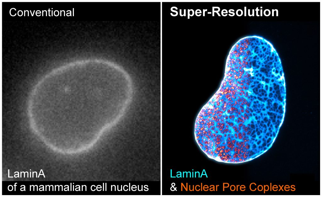
Seeing is believing. A major objective of our research program is to directly visualize how cells organize proteins, RNA, and DNA in space and time. We have developed a series of super-resolution microscopy methods to dissect the architecture of cellular structures at molecular resolution and with high labeling efficiency, brightness, and detection sensitivity. We are particularly interested in the structure-function relationships of the nuclear lamina, chromatin, cytoplasmic vesicles, lysosomes, and cellular protrusions called cilia.
Methods: Expansion Microscopy (ExM), Stochastic Optical Reconstruction Microscopy (STORM), Structured Illumination Microscopy (SIM), Airyscan Microscopy, live-cell imaging
II. How single-cell multiomic data are organized at the spatial scale?

Over the last few years, we are witnessing rapid advances in the fields of single-cell transcriptomics, genomics, and proteomics. However, most tissues and organs are solid, with cells encased within a complex extracellular matrix network. Studying single cells from solid tissues usually requires their mechanical and enzymatic disaggregation. As a result, the positional information is lost, which is otherwise essential for a proper understanding of how cells cooperate in tissues. We take optical and chemical approaches to develop novel, spatially-resolved multiomic methods, which isolate cells without homogenization, enable cell phenotyping with super-resolution, and allow multiomics in the same single cells. This novel platform will significantly advance our knowledge of how cell phenotypes are maintained in native tissue and become perturbed in disease.
III. New and better biomarkers for diagnosis and precision medicine
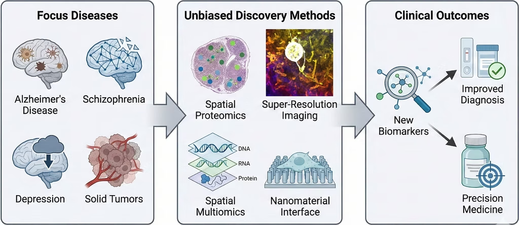
We are dedicated to discovering new and better biomarkers to advance diagnosis and precision medicine related to aging, including Alzheimer’s disease, schizophrenia, depression, and solid tumors. By employing unbiased discovery methods developed in our lab, such as spatial proteomics, spatial multiomics, super-resolution imaging, and nanomaterial interfaces, we aim to identify novel biomarkers that directly lead to improved diagnostic tools and more effective, personalized precision medicine strategies.
Awards and Honors
National Alzheimer’s Coordinating Center (NACC) Rising Star Award, 2025
NSF CAREER Award, NSF, 2024-2029
NIH Director’s New Innovator Award (DP2), NIH,2022-2027
Chan Zuckerberg Initiative Advancing Imaging Through Collaborative Projects, Chan Zuckerberg Initiative, 2023-2026
Hellman Fellow, Hellman Foundation, 2022-2023
Chan Zuckerberg Initiative Visual Proteomics Imaging Award, Chan Zuckerberg Initiative, 2021-2024
NIH Pathway to Independence Award (K99/R00), NIGMS,2018-2023
Recent Publications
- Y. Zhuang, Z. Zhang, Z. Dai, X. Shi#, Landscape Expansion Microscopy Reveals Interactions between Membrane and Phase-Separated Organelles, Journal of Cell Biology, 225, e202502035, 2026. **Image on Table of Contents**
- X. Zhao#, V. Mouilleau, Y. Wang, A.C. Solak, J.Q. Garcia, X. Chen, X. Shi, C.J. Wilkinson, L.A. Royer#, Z. Dong#, S. Guo#, PCM1 coordinates centrosome asymmetry with polarized endosome dynamics to regulate daughter cell fate, Nature Communications, 16, 10728, 2025.
- J.A. Woytash, A.E.Y.T. Lefebvre, Z. Zhang, B. Xu, S.A. Harchenko, H.T. Le, A.R. McColloch, X. Shi, M.A. Digman, & O.V. Razorenova, CDCP1/mitochondrial Src axis increases electron transport chain function to promote metastasis in triple-negative breast cancer, British Journal of Cancer, 133, 1265–1277, 2025.
- E.G. Stokes, J.J. Vasquez, G. Azouz, M. Nguyen, A. Tierno, Y. Zhuang, V.M. Galinato, M. Hui, M. Toledano, I. Tyler, X. Shi, R.F. Hunt, J. Aoto, K.T. Beier, Cationic peptides cause memory loss through endophilin-mediated endocytosis, Nature, 638, 479-489, 2025.
- Y. Yoon, E. Bournique, L.V. Soles, H. Yin, H.-F. Chu, C. Yin, Y. Zhuang, X. Liu, L. Liu, J. Jeong, C. Yu, M. Valdez, L. Tian, L. Huang, X. Shi, G. Seelig, F. Ding, L. Tong, R. Buisson, Y. Shi, RBBP6 anchors pre-mRNA 3′ end processing to nuclear speckles for efficient gene expression, Molecular Cell, 85, 555, 2025.
- S. Park, X. Wang, Y. Mo, S. Zhang, X. Li, K.C. Fong, C. Yu, A.A. Tran, L. Scipioni, Z. Dai, X. Huang, L. Huang, X. Shi#, Proximity Labeling Expansion Microscopy (PL-ExM) resolves structure of the interactome, Journal of Materials Chemistry B, 12, 8335, 2024.
Back Cover image - Y. Zhuang, X. Shi#, Label-Retention Expansion Microscopy (LR-ExM) for Enhanced Fluorescent Signals using Trifunctional Probes, Current Protocols 4, e973, 2024.
- Y. Zhuang, X. Shi#, Expansion microscopy: A chemical approach for super-resolution microscopy, Current Opinion in Structural Biology 81, 102614, 2023.
- S. Park, X. Shi#, Expansion microscopy of ciliary proteins, Cilia Methods and Protocols, Methods in Molecular Biology, Chapter 4, Springer Nature, 2023.
- S. Park, Y. Zhuang, X. Shi#, Label-retention expansion microscopy (LR-ExM) enables super-resolution imaging and high-efficiency labeling, the Journal of Visualized Experiments (JoVE), e63793, 2022. [link]Label-retention expansion microscopy, X. Shi * #, Q. Li *, Z. Dai, A.A.Tran, S. Feng, A.D. Ramirez, Z. Lin, X. Wang, T.T. Chow, J. Chen D. Kumar, A. McColloch, J.F. Reiter, E.J. Huang, I.B. Seiple #, B. Huang #,
J. Cell Biol. 220 (9): e202105067 (2021). **Image on Table of Content Page** - Polarized endosome dynamics engage cytoplasmic Par-3 that recruits dynein during asymmetric cell division. X. Zhao, J.Q. Garcia, K. Tong, X. Chen, B. Yang, Q. Li, Z. Dai, X. Shi, I.B. Seiple, B. Huang, S. Guo,
Science Advance, 7, eabg1244 (2021) [link] - Kinase-mediated RAS signaling via membraneless cytoplasmic protein granules, A. Tulpule, J. Guan, D.S. Neel*, H.R. Allegakoen, Y.P. Lin, D. Brown, Y.-T. Chou, A. Heslin, N. Chatterjee, S. Perati, S. Menon, T.A. Nguyen, J. Debnath, A.D. Ramirez, X. Shi, B. Yang, S. Feng, S. Makhija, B. Huang#, T.G. Bivona#,
Cell, 184, 2649 (2021) [link] - A ciliopathy complex builds distal appendages to initiate ciliogenesis, D. Kumar, A. Rains, V. Herranz-Pérez, Q. Lu, X. Shi, D.L. Swaney, E. Stevenson, N.J. Krogan, B. Huang, C. Westlake, J.M. Garcia-Verdugo, B. Yoder, J.F. Reiter,
J. Cell Biol. 220 (9): e202011133 (2021). [link] - Neurotoxic microglia promote TDP-43 proteinopathy in progranulin deficiency, J. Zhang, D. Velmeshev, K. Hashimoto, Y.-H. Huang, J. W. Hofmann, X. Shi, J. Chen, A. M. Leidal, J. G. Dishart, M. K. Cahill, K. W. Kelley, S. A. Liddelow, W. W. Seeley, B. L. Miller, T. C. Walther, R. V. Farese Jr., J. P. Taylor, E. M. Ullian, B. Huang, J. Debnath, T. Wittmann, A. R. Kriegstein & E. J. Huang,
Nature, 588, 459 (2020) [link] - Nanotopography enhances dynamic remodeling of tight junction proteins through cytosolic complexes, X. Huang, X. Shi, M.E. Hansen, C.L. Nemeth, A. Ceili, B. Huang, T. Mauro, M. Koval, T.A. Desai,
ACS Nano, 14, 13192 (2020). [link] - X. Shi, G. Garcia, Y. Wang, J. Reiter, B. Huang, Alignment of super-resolution images for semi-flexible structures in 3D, PLoS One 0212735 (2019). [link]
- T.T. Chow, X. Shi, J.H. Wei, J. Guan, G. Stadler, B. Huang, E. Blackburn, Local enrichment of HP1alpha at telomeres alters their structure and regulation of telomere protection,
Nature Communications 9, 3583 (2018). [link] - X. Shi*, G. Garcia*, J.C. Van De Weghe, R. McGorty, G.J. Pazour, D. Doherty, B. Huang#, J.F. Reiter#, Super-resolution microscopy reveals that disruption of ciliary transition zone architecture is a cause of Joubert syndrome,
Nature Cell Biology 19, 1178 (2017). [link] **cover article** - J. Guan, H. Liu, X. Shi, S. Feng, B. Huang, Tracking multiple genomic elements using correlative CRISPR imaging and sequential DNA FISH,
Biophysical Journal 112,1077 (2017). [link] **New and Notable**