Thomas Schilling, Ph.D.

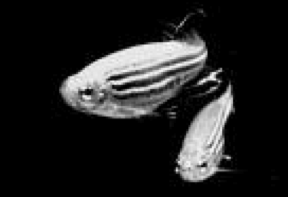
Zebrafish developmental genetics – Our laboratory uses genetics and molecular biology to study pattern formation in the musculoskeletal and nervous systems of early zebrafish embryos. Their rapid development and simple anatomy, together with recently developed techniques for reverse genetics and a complete genome sequence, make zebrafish a powerful molecular genetic system for studying the mechanisms of development. We focus on several areas: (i) formation of the head skeleton as a model for human craniofacial birth defects and (ii) cell fate determination and polarity in migratory cells that form cranial cartilage, muscle and tendon, and 3) patterning of the nervous system, particularly the brainstem that innervates these craniofacial structures and controls behaviors such as feeding and breathing. We also have close collaborations with labs studying development and physiology of the lens, where we have developed zebrafish models for human congenital cataract. In all cases, we are interested in how gene functions translate into cell behaviors and the formation of tissues and organs.

A major focus of the work in our laboratory is to define as fully as possible the genetic and molecular pathways that establish an embryonic population of migratory cells called the neural crest (NC) and its derivatives. NC cells get their name from the fact that they arise from the crests of the forming neural tube as it folds, and give rise to all of the body’s pigmentation, most of its peripheral nervous system, as well as the head skeleton. We have analyzed and cloned genes underlying a large library of mutations that affect craniofacial development. We have also identified genes that control NC migration, and these share many similarities with other migrating cells, such as metastatic cancer cells.
A recent focus of our lab is to understand the genetic and molecular pathways that specify cell fates using single cell transcriptomics, including NC cells. Here we combine experiments with a more computational, systems approach. These studies have identified distinct transitional states in NC cells as they first acquire different identities as well as the temporal dynamics of signals involved in regulating these transitions such as Wnts. Single cell studies of one very intriguing NC derivative, the tendon fibroblast (tenocyte) are now revealing unexpected heterogeneity and responses to mechanical force in this population, which have implications for therapeutic for tendon injuries and diseases (tendinopathies).
Recent Publications
- Vorontsova I, Vallmitiana A, Torrado B, Schilling TF, Hall JE, Gratton E and Malacrida L (2022). In vivo macromolecular crowding is differentially modulated by aquaporin 0 in zebrafish lens: insights from a nanoenvironment sensor and spectral imaging. Science Advances Feb 18;8(7):eabj4833. doi: 10.1126/sciadv.abj4833.
- Hoyle D, Dranow D and Schilling TF (2022). Pthlha and mechanical force control early patterning of growth zones in the zebrafish craniofacial skeleton. Development Jan 15;149(2):dev199826. doi/10.1242/dev.199826.
- Tatarakis D, Cang Z, Wu X, Karikomi M, MacLean A, Nie Q and Schilling TF (2021). Single cell transcriptomic analysis of zebrafish cranial neural crest reveals spatiotemporal regulation of lineage decisions during development. Cell Reports Dec 21;37(12):110140. doi: 10.1016/j.celrep.2021.110140.
- Vorontsova I, Hall JE, Schilling TF, Nagai N and Nakazawa Y (2021). Differences in a single extracellular residue underlie adhesive functions of two zebrafish Aqp0s. Cells 6;10(8):2005. doi: 10.3390/cells10082005.
- Qiu Y, Fung L, Schilling TF# and Nie Q# (2021). Multiple morphogens and rapid elongation promote segmental patterning during development. PLoS Computational Biology 17(6):e1009077. doi: 10.1371/journal.pcbi.1009077.. #co-corresponding authors
- Reynolds S, Pierce C, Powell B, Kite A, Hall-Ruiz N, Schilling TF and Le Pabic P (2021). A show of Hands: novel and conserved expression patterns of teleost Hand orthologues during craniofacial, heart, fin, peripheral nervous system and gut development. Developmental Dynamics 2021 Jun 6. doi: 10.1002/dvdy.380.
- Wang K, Vorontsova I, Hoshino M, Uesugi K, Yagi N, Hall JE, Schilling TF and Pierscionek BK (2021). Aquaporins have regional functions in development of refractive index in the zebrafish eye lens. Investigative Opthalmology and Visual Science March 2021 doi:https://doi.org/10.1167/iovs.62.3.23.
- Heubel B, Bredesen C, Schilling TF and LePabic P (2020). Endochondral growth zone pattern and activity in the zebrafish pharyngeal skeleton. Developmental Dynamics 2020 Aug 27 doi:10.1002/dvdy.241.
- Niu X, Subramanian A, Hwang TH, Schilling TF and Galloway JL (2020). Tendon cell regeneration is mediated by attachment-site resident progenitors and BMP signaling. Current Biology 2020 Jun 29:S0960-9822(20)30832-0. doi: 10.1016/j.cub.2020.06.016
- Wang K, Vorontsova I, Hoshino M, Uesugi K, Yagi N, Hall JE, Schilling TF and Pierscionek BK (2020). Optical development in the zebrafish eye lens. FASEB Journal doi.org/10.1096/fj.201902607R
- Hendrick L, Carter GA, Hilbrands EH, Heubel BP, Schilling TF and Le Pabic P (2019). Tracking pigment progenitors reveals distinct modes of bar, stripe and spot development in sand-dweller cichlids from Lake Malawi. EvoDevo 2019 Aug 12;10:18. doi: 10.1186/s1186/s13227-019-0132-7.
- Vorontsova I, Hall J and Schilling TF (2019). Assessment of zebrafish lens nucleus localization and sutural integrity. Journal of Visualized Experiments (JoVE) May 6;(147). doi:10.3791/59528.
- Sharma P, MacLean A, Clouthier D, Nie Q and Schilling TF (2019). Transcriptomics reveals complex kinetics of dorsal-ventral patterning gene expression in the mandibular arch. Genesis 2018 Dec 18:e23275. doi:10/1002/dvg.23275.
- Subramanian A, Kanzaki L, Galloway JL and Schilling TF (2018). Mechanical force regulates tendon extracellular matrix organization and tenocyte morphogenesis through TGFbeta signaling. eLife 2018;7. E38069. doi: 10.7554/eLife.38069.
- Meinecke L*, Sharma P*, Du H, Zhang L, Nie Q# and Schilling TF# (2018). Modeling craniofacial development reveals spatiotemporal constraints on robust patterning of the mandibular arch. PLoS Computational Biology 14(11):e1006569. doi: 10.1371/journal.pcbi.1006569. (*co-first authors; # – co-senior authors)
- Vorontzova I, Gehring I, Hall JE, and Schilling TF (2018). Aquaporin-0 regulates suture stability in the zebrafish lens. Investigative Ophthalmology and Visual Science 59, 2869-2879. doi:10.1116/iovs.18-24044.
- Rackaukas C, Schilling TF and Nie Q (2018). Mean-independent noise attenuation via intermediate states. iScience 3, 11-20. https:oi.org/10.1016/j.isci.2018.04.002.
- Aguillon R, Batut J, Subramanian A, Madelaine R, Dufourcq P, Schilling TF and Blader P (2018). Cell-type heterogeneity in the zebrafish olfactory placode is generated from progenitors within the preplacodal ectoderm. eLife 2018;7:e32041: doi: https://doi.org/org/10.7554/eLife.32041.
- Wang W, Holmes WR, Sosnik J, Schilling TF and Nie Q (2017). Cell sorting and noise-induced plasticity coordinate to sharpen boundaries between gene expression domains. PLoS Computational Biology 13(1):e1005307. doi: 10.1371/journal.pcbi.1005307.
- Muto A and Schilling TF (2017). Zebrafish as a model to study cohesin and cohesinopathies. In K Yokomori and Katsuhiko Shirahige (Eds.). Cohesin and Condensin: Methods and Protocols. Methods in Mol Biol. Vol 1515, pp. 177-196 Springer Press.
- Vibert L, Aquino G, Gehring I, Subkhankulova T, Schilling TF, Rocco A and Kelsh RN (2016). An ongoing role for Wnt signaling in differentiating melanocytes in vivo. Pigment Cell Melanoma Research doi: 10.1111/pcmr.12568.
- Nichols J, Dowd J, Watson S, Parthasarathy R, Brooks E, Subramanian A, Nachtrab G, Poss KD, Schilling TF, and Kimmel CB. (2016). Repetitive element silencing buffers a mef2ca dependent ligament versus bone fate decision in the zebrafish craniofacial skeleton. Development 143, 4430-4440.
- Schilling TF, Sosnik J and Q Nie (2016). Visualizing retinoic acid morphogen gradients. In HW Detrich III, M Westerfield and LI Zon (Eds.). The Zebrafish: Cellular and Developmental Biology, Part A Cellular Biology (pp. 139-163). Methods in Cell Biol Vol 133. Elsevier Inc., Academic Press.
- Kawauchi S, Santos R, Muto A, Lopez-Burks M, Schilling TF, Lander AD and Calof AL (2016). Using vertebrate animal models to understand the etiology of developmental defects in Cornelia de Lange Syndrome. Am J Hum Genet C Semin Med Genet doi:10.1002/ajmg.c.31484.
- Sosnik J, Zheng L, Rackauckas CV, Gratton E, Nie Q, and Schilling TF. (2016). Noise modulation of retinoic acid signaling controls sharpening of segmental gene expression boundaries in the zebrafish hindbrain. eLife 5:e14034. doi:10.7554/eLife.14034.
- Le Pabic P, Cooper J and Schilling TF (2016). Developmental basis of phenotypic integration in two Lake Malawi cichlids. EvoDevo 21 7:3 doi: 10.1186/s13227-016-0040-z.
- Subramanian A, and Schilling TF (2015). Tendon development and musculoskeletal assembly: emerging roles for the extracellular matrix. Development 142, 4191-4204. doi: 10.1242/dev.114777.
- Cruz IA, Kappedal R, Mackenzie S, Hoffman TL, Schilling TF and Raible DW (2015). Robust regeneration of adult zebrafish lateral line hair cells reflects continued precursor pool maintenance. Developmental Biology 402, 229-238. doi: 10.1016/j.ydbio.2015.03.019.
- Boer, EF, Howell, ED, Schilling, TF, Jette, CA and Stewart RA (2015). Fascin1-dependent filopodia are required for directional migration of a subset of neural crest cells. PLoS Genetics 11(1):e1004946. doi: 10.1371/journal.pgen.1004946.
- Le Pabic P, Ng C and Schilling TF (2014). Fat-Dachsous signaling coordinates cartilage differentiation and polarity during craniofacial development. PLoS Genetics 10(10):e1004726. doi: 10.1371/journal.pgen.1004726.
- Muto A, Ikeda S, Lopez-Burks ME, Kikuchi Y, Calof AL, Lander AD and Schilling TF (2014). Nipbl and Mediator cooperatively regulate gene expression to control limb development. PLoS Genetics 10(9):e1004671. doi: 10.1371/journal.pgen.1004671.
- Alexander C, Piloto S, Le Pabic P and Schilling TF (2014). Wnt signaling interacts with Bmp and Edn1 to regulate dorsal-ventral patterning and growth of the craniofacial skeleton. PLoS Genetics 10(7):e1004479. doi: 10.1371/journal.pgen.1004479.
- Subramanian A and Schilling TF (2014). Thrombospondin-4 controls matrix assembly during development and repair of myotendinous junctions. eLife (Cambridge) doi: 10.7554/eLife.02372.
- Tuttle A, Hoffman T and Schilling TF (2014). rabconnectin-3a regulates vesicle endocytosis and canonical Wnt signaling in zebrafish neural crest migration. PLoS Biology 12(5): e1001852. doi:10.1371/journal.pbio.1001852. Article recommended in Faculty of 1000.
- Schilling TF and Le Pabic P (2014). Neural crest cells in craniofacial skeletal development. In Neural Crest Cell Differentiation and Disease (ed. P Trainor). (pp. 127-151). Elsevier.


